
Cell therapy based on human pluripotent stem cells (hPSCs) is showing great promise, with 205 clinical studies underway worldwide (source: https://hpscreg.eu/) at time of writing. Japan has also seen major breakthroughs, with two commercialized products: ReHeart developed by Cuorips Inc. and Amchepry by Sumitomo Pharma Co. and Racthera Inc. available on the market since February 2026. Several other promising candidates are progressing through various stages of development, including Bluerock’s Bendaneprocel, Vertex’s Zimislecel, and Gameto’s Fertilo system, all of which have reached Phase 3.
These are exciting times, but the emergence of genetic variants during extensive in-vitro culture remains a significant challenge for scientists working on these cell types. Such genetic defects are an unavoidable byproduct of the development, growth, and differentiation of hPSCs for therapeutic applications, and they cannot be ignored: they risk reducing the efficacy of therapies and, more concerning still, pose a safety risk given the potential tumorigenic properties of variant cells. They can also compromise basic research, since experimental work with variant hPSCs, which may show aberrant proliferation rates or altered differentiation capacity, can generate unpredictable results, undermining the reliability of stem cell-based disease modeling and developmental studies (Andrews, 2021).
This is why it is essential for iPSC scientists to establish a robust genomic stability workflow to derisk their iPSC cultures.
With over 8 years of experience working with scientists around the world, Stem Genomics has developed an effective strategy for the early detection of genomic variants in hPSCs. Before presenting this strategy, let’s take a look at the variants you’re likely to encounter.
The genomic variants encountered in human pluripotent stem cell cultures
They fall into two distinct categories, with one subcategory:
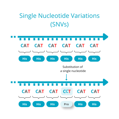
- Single Nucleotide Variations (SNVs): SNVs occur when a single nucleotide (adenine, thymine, cytosine, or guanine) in the genome sequence is substituted, with stop-gain variants being the most likely to cause disease. They are closely linked to a range of physiological and pathological processes and can serve as important biomarkers. Consequently, accurate and sensitive identification of SNVs is critical for medical diagnosis and disease treatment. SNVs are very small, by definition, just 1 base pair in size, which makes their detection technically demanding.
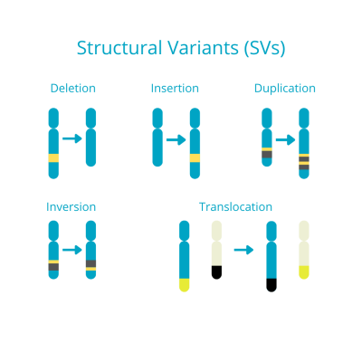
- Structural variants (SVs)/Copy Number Variations (CNVs): SVs are large-scale karyotypic changes ranging in size from 50 base pairs to several million base pairs. They can create novel breakpoint junctions associated with normal genomic variation, Mendelian diseases, or complex disease traits, and they contribute significantly to overall genomic diversity. SVs can take the form of simple genomic rearrangements, such as copy-number variations (CNVs, deletions or duplications), or complex genomic rearrangements (CGRs), classified by the number of breakpoint junctions involved, with CGRs defined as having more than two (e.g., duplication–normal–duplication, duplication–triplication/inversion duplication) (Pande et al., 2025). SVs have only recently come under closer study, thanks to advanced technologies such as NGS that have made it possible to capture them more effectively. CNVs, which typically range from 1,000 base pairs to 5 megabases, the resolution level of cytogenetic analysis, are now recognized as a major source of structural variation in the genome. Evidence continues to accumulate that CNVs play an important role in human disease (source: https://www.nature.com/scitable/topicpage/copy-number-variation-and-human-disease-741737/). Last but not least, CNVs are, in fact, the most common recurrent abnormality observed in human pluripotent stem cells. This is a key characteristic that has led Stem Genomics to develop its own proprietary CNV database, used in its targeted digital-PCR and NGS-based detection solutions (Assou et al., 2020).
The recurrence of genomic variants found in hPSCs
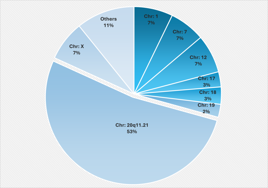
Several studies have identified that genomic variants in iPSCs and before them, in ESCs, tend to be recurrent (Assou et al., 2020; Draper et al., 2004; Lefort et al., 2008; Spits et al., 2008). Specific amplifications on chromosomes 1, 12, 17, and 20 are frequently observed, with gains in the 20q11.21 region being particularly common in hPSC lines (Figure 1) (Maitra et al., 2005; (Amps et al., 2011)). These recurrent variants were brought to light through methods offering finer resolution than traditional G-banding karyotyping, such as CGH, SNP arrays, and digital PCR.
More recently, whole genome and exome sequencing, alongside analysis or RNAseq data has identified frequent single nucleotide variations (SNVs) in hPSCs. Even at the SNV level, a common set of variants appears to recur, particularly in cancer-related genes such as Tumor Protein P53 (TP53) and BCL6 Corepressor (BCOR). (Vales & Barbaric, 2024)
Figure 1: Localization of the most frequent genomic abnormalities observed in Stem Genomics testing using digital PCR
This figure illustrates the distribution of genetic abnormalities in human pluripotent stem cells (hPSCs) across different chromosomes as detected by digital droplet PCR. The most affected region was 20q11.21, followed by chromosomes 12, 7, 1 and X, which showed similar patterns. The graph underscores the importance of regular genomic monitoring to maintain the integrity of hPSCs for both research and clinical applications. The data, derived from 2,124 abnormal samples, identified 2,374 chromosomal abnormalities, as some samples had multiple alterations. This proprietary data was provided by Stem Genomics. (Source: Stem Genomics White Paper “Ensuring genetic integrity in human Pluripotent Stem Cells – Challenges and solutions”, 2024)
A proposed strategy for optimum genomic abnormalities detection
The schematic figure below shows, at a glance, the key stages at which Stem Genomics recommends testing hPSCs in culture for genetic variants. This strategy has been designed to be practical for most labs and follows several recommendations outlined in the ISSCR Standards for Human Stem Cell Use in Research and the FDA’s Safety Testing of Human Allogeneic Cells Expanded for Use in Cell-Based Medical Products guidelines.
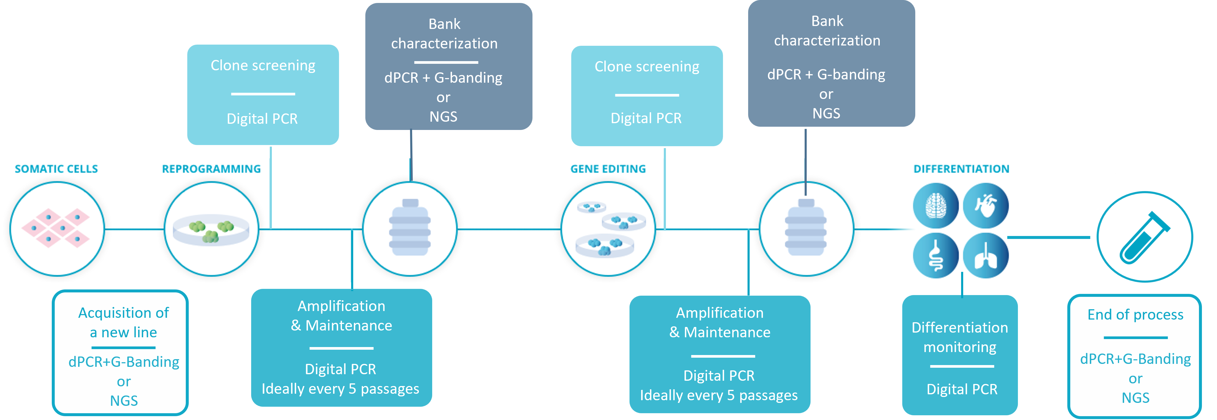
The workflow for hPSC genetic integrity quality control recommended by Stem Genomics.
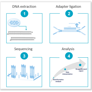
- Acquisition of a new line: Before starting any work, it is critical to confirm the genomic stability of the initial material. Most purchased lines come with some form of genomic stability testing, but we have encountered several instances of small abnormalities that went undetected by G-banding karyotyping alone. We therefore recommend pairing G-banding with digital PCR or, budget permitting, running NGS testing*, which is also an excellent way to establish an initial mutational profile for your cell lines, one you can refer back to later on.
- Reprogramming and gene editing: These procedures can favor the generation and selection of genomic aberrations in PSCs. Quick screening of clones and colonies using digital PCR is possible and essential before any subsequent use.
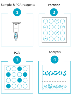
- In-process control during cell amplification & maintenance: This is another stress factor likely to generate genetic defects in hPSCs. Given the speed at which a recurrent abnormality can take over a culture, Stem Genomics and others (McIntire et al., 2020) recommend testing PSCs at least every 5–10 passages. In practice, this is only feasible using digital PCR, since its multiplexing capabilities allow broad detection coverage with a fast turnaround and affordable cost.
- Pre-banking characterization: Genomic stability is one of the critical features required for the complete characterization of a cell line before banking. For this stage, we offer two different methods, and the choice between them is often budget-dependent.
- Differentiation monitoring: We recommend continuing to check for genomic abnormalities until your cells have reached full differentiation, testing each time you change the media. Once again, the speed of digital PCR makes it ideal for this stage.
- End of process: A final check will be required before publication, as well as before advancing to the clinical stage. If you performed an NGS* test at the start of the process, it is good practice to repeat it at the end in order to evaluate the impact of the culture process on the baseline genetic landscape. If cost is prohibitive, an alternative is to combine G-banding karyotyping with digital PCR, which offers standard structural rearrangement analysis alongside digital PCR’s resolution, capable of detecting sub-karyotypic abnormalities such as 20q.
*Please note that the FDA guidelines “Safety Testing of Human Allogeneic Cells Expanded for Use in Cell-Based Medical Products guidelines” recommend a Whole Genome Sequencing screening at a minimum depth of 50x to ensure the safety of cellular products.
- More information on digital PCR
- More information on Next Generation Sequencing
Sources:
Amps, K., Andrews, P. W., Anyfantis, G., Armstrong, L., Avery, S., Baharvand, H., Baker, J., Baker, D., Munoz, M. B., Beil, S., Benvenisty, N., Ben-Yosef, D., Biancotti, J. C., Bosman, A., Brena, R. M., Brison, D., Caisander, G., Camarasa, M. V., Chen, J., … Zhou, Q. (2011). Screening ethnically diverse human embryonic stem cells identifies a chromosome 20 minimal amplicon conferring growth advantage. Nature Biotechnology, 29(12), 1132–1144. https://doi.org/10.1038/nbt.2051
Andrews, P. W. (2021). Human pluripotent stem cells: Genetic instability or stability? In Regenerative Medicine (Vol. 16, Number 2, pp. 113–115). Future Medicine Ltd. https://doi.org/10.2217/rme-2021-0013
Assou, S., Girault, N., Plinet, M., Bouckenheimer, J., Sansac, C., Combe, M., Mianné, J., Bourguignon, C., Fieldes, M., Ahmed, E., Commes, T., Boureux, A., Lemaître, J. M., & De Vos, J. (2020). Recurrent Genetic Abnormalities in Human Pluripotent Stem Cells: Definition and Routine Detection in Culture Supernatant by Targeted Droplet Digital PCR. Stem Cell Reports, 14(1), 1–8. https://doi.org/10.1016/j.stemcr.2019.12.004
Draper, J. S., Smith, K., Gokhale, P., Moore, H. D., Maltby, E., Johnson, J., Meisner, L., Zwaka, T. P., Thomson, J. A., & Andrews, P. W. (2004). Recurrent gain of chromosomes 17q and 12 in cultured human embryonic stem cells. Nature Biotechnology, 22(1), 53–54. https://doi.org/10.1038/nbt922
Lefort, N., Feyeux, M., Bas, C., Féraud, O., Bennaceur-Griscelli, A., Tachdjian, G., Peschanski, M., & Perrier, A. L. (2008). Human embryonic stem cells reveal recurrent genomic instability at 20q11.21. Nature Biotechnology, 26(12), 1364–1366. https://doi.org/10.1038/nbt.1509
Spits, C., Mateizel, I., Geens, M., Mertzanidou, A., Staessen, C., Vandeskelde, Y., Van Der Elst, J., Liebaers, I., & Sermon, K. (2008). Recurrent chromosomal abnormalities in human embryonic stem cells. Nature Biotechnology, 26(12), 1361–1363. https://doi.org/10.1038/nbt.1510
Vales, J. P., & Barbaric, I. (2024). Culture-acquired genetic variation in human pluripotent stem cells: Twenty years on. In BioEssays (Vol. 46, Number 12). John Wiley and Sons Inc. https://doi.org/10.1002/bies.202400062